Immunohistochemical Profile of Renal Cell Carcinoma in Patients Younger than 45 Years of Age: Analysis of 87 Cases of Different Tumor Subtypes
Ryan Carr¹, Guoping Cai¹, Yunhua Bao¹, Manju L Prasad¹, Satish K Tickoo2, Adebowale J. Adeniran
1Department of Pathology, Yale School of Medicine, New Haven, CT; 2Memorial Sloan-Kettering Cancer Center, New York, NY, USA
ABSTRACT
Background:
Renal cell carcinoma (RCC) is distinctly rare in children and young adults. Because of the rarity of the tumor in this age group, the immunohistochemical profile has not been fully described. The goal of this study is to define the immunohistochemical profile of the various subtypes of RCC in patients aged 45 years and younger.
Design:
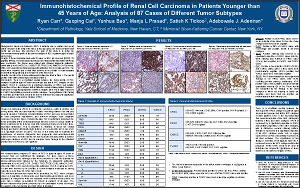
A tissue microarray block with 87 cases of RCC (49 clear cell [CRCC], 23 papillary [PRCC], 10 chromophobe [ChRCC] and 5 translocation-associated [TxRCC]) was constructed. The tumors were stained with following diagnostic antibodies: cytokeratin (CK) AE1/AE3, CK7, CK903, TFE3, TFEB, CD10, AMACR, C-KIT, vimentin, EMA, CAIX; and with the following prognostic antibodies: PAX-8, survivin and VHL. The slides were reviewed and a stain was deemed to be positive if it demonstrated moderate to strong intensity in more than 5% of the tumor cells.
Results:
Results of the immunohistochemical stains are listed in Table 1. The overall profiles of the different tumor types are listed in Table 2.
Conclusion:
There is a significant overlap between the immunoprofile of RCC subtypes in patients aged 45 years and younger and that in RCC subtypes of older patients. PAX-8 is expressed by a significant proportion of PRCC and variable proportions of the other subtypes, with a distinctive nuclear staining pattern. Survivin is expressed in a significant proportion of all RCC subtypes while VHL is highly expressed in ChRCC and variably expressed in all other subtypes. The significance of this finding is uncertain, as further work needs to be done to determine the prognostic significance of these markers on the various subtypes of RCC.
©2012 Yale Department of Pathology. All rights reserved.
Any redistribution or reproduction of part or all of the contents in any form is prohibited. You may not, except with express written permission of the author or the Department of Pathology, distribute or commercially exploit the content, nor may you transmit it or store it in any other website or other form of electronic retrieval system, including use for educational purposes.