Prognostic Significance of PAX8, Survivin, and Ki-67 Expression in Pancreatic Endocrine Neoplasms: A Tissue Microarray Analysis
Jane Bernstein, M.D., Alison L. Van Dyke, M.D., Ph.D., Yunhua Bao, M.D., Fang Bao, M.D., Barton Kenney, M.D., Manju Prasad, M.D., Guorong Chen, M.D., Marie Robert, M.D., Guoping Cai, M.D.
Department of Pathology, Yale School of Medicine, New Haven, CT, USA
ABSTRACT
Pancreatic endocrine neoplasms (PENs) are are relatively uncommon tumors of the pancreas, which account for less than 2% of of all pancreatic neoplasm. PENs can be sporadic or associated with genetic syndromes including multiple endocrine neoplasia type 1, von Hippel-Lindau disease, von Recklinghausen disease, and tuberous sclerosis. They are typically seen in middle-age adults and are divided into functional and non-functional tumors.
Accurate classification/grading of PENs is important for appropriate clinical management and prognosis prediction of the patients. According to the current WHO classification, PENs include well-differentiated endocrine tumor with benign behavior, well-differentiated endocrine tumor with uncertain behavior, well-differentiated endocrine carcinoma, and poorly differentiated carcinoma. This classification is based on the presence or absence of local invasion, lymph node or distant metastasis, as well as expression of Ki-67. There are, however, no widely-accepted immunohistochemical markers that can reliably grade the tumors and predict tumor behavior.
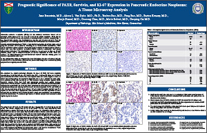
In this retrospective study, we assessed the potential prognostic value of markers that are associated with cell proliferation, differentiation, and survival using a tissue microarray (TMA) prepared from surgically resected PENs.
©2012 Yale Department of Pathology. All rights reserved.
Any redistribution or reproduction of part or all of the contents in any form is prohibited. You may not, except with express written permission of the author or the Department of Pathology, distribute or commercially exploit the content, nor may you transmit it or store it in any other website or other form of electronic retrieval system, including use for educational purposes.