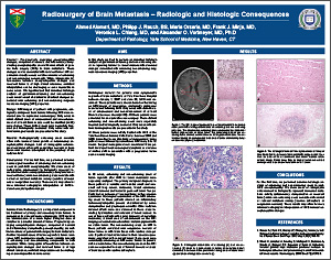
Radiosurgery of Brain Metastasis – Radiologic and Histologic Consequences
Ahmed Alomari, MD, Philipp J. Rauch, BS, Maria Orsaria, MD, Frank J. Minja, MD, Veronica L. Chiang, MD, and Alexander O. Vortmeyer, MD, Ph.D
Department of Pathology, Yale School of Medicine, New Haven, CT, USA
ABSTRACT
Context: Progressively enlarging encephalopathic changes, composed of necrosis and inflammatory demy-elinating encephalopathy, are well-known effects of gam-ma knife surgery (GKS) to brain metastasis. These changes can be associated with recurrent tumor. MRI ex-amination usually reveals variable amounts of enhancing and non-enhancing components. While radiographic dif-ferentiation between encephalopathic changes and recurrent tumor is of high clinical relevance, confident interpretation can be challenging or even impossible in some cases. We hypothesized that individual histologic analysis of gadolinium-enhancing and non-enhancing areas would reveal characteristic structural changes as-sociated with enhancing and non-enhancing magnetic resonance imaging (MRI) properties.
Design: MRI-images of patients with progressive, etio-logically ambiguous brain lesions following GKS were re-viewed prior to explorative neurosurgery. Only cases in which distinct areas of enhancement and non-enhance-ment of at least 5mm in size could be identified (n=18) were chosen. These distinct areas were separately biop-sied and histologically evaluated. Only cases with uni-form histological results are presented in this study.
Results: Radiographically enhancing areas correlate either with recurrent tumor growth or inflammatory en-cephalopathic changes. Lack of radiographic enhance-ment correlates either with coagulative necrosis or brain tissue with reactive astrocytosis in the periphery of the radiation field.
Conclusions: For the first time, we performed selective neurosurgical resection of enhancing and non-enhancing areas in post-GKS encephalopathy. We show areas of radiographic enhancement to biologically represent in-creased metabolic activity (inflammatory demyelination or recurrent tumor) while non-enhancing areas correlate with normal or reduced metabolic activity (reactive astrocyt-osis or coagulation necrosis). These results may allow for more informed radiographic interpretation of GKS-in-duced encephalopathic changes.
©2012 Yale Department of Pathology. All rights reserved.
Any redistribution or reproduction of part or all of the contents in any form is prohibited. You may not, except with express written permission of the author or the Department of Pathology, distribute or commercially exploit the content, nor may you transmit it or store it in any other website or other form of electronic retrieval system, including use for educational purposes.