Comparison of Immunohistochemical Profiles of Papillary Renal Cell Carcinoma Subtypes
Ahmed Alomari1, Harriet Kluger2, Kenneth Haines1, Dinesh Singh3, Manju L. Prasad1, Adebowale J. Adeniran1
Departments of 1Pathology, 2Medical Oncology, and 3Urology, Yale School of Medicine, New Haven, CT, USA
ABSTRACT
Background: Papillary renal cell carcinoma (PRCC) is characterized morphologically by the presence of fibrovascular cores, lined by papillae of malignant epithelial cells. It is further subclassified into 2 types based on cell type. Type 2 tumors are generally believed to have a poorer prognosis than Type 1 tumors. This prognostic difference has led some to believe that some of the Type 2 tumors may actually represent a separate entity from PRCC. This study was designed to compare the immunohistochemical profiles in both subtypes of PRCC.
Design: A tissue microarray block with 124 cases of PRCC was constructed. The tumors were stained with the following diagnostic antibodies: CK7, EMA, CD10, CAIX, vimentin, CK903, C-kit, racemase, PAX-8, PAX-2 and CEA. A stain was deemed to be positive if it demonstrated at least weak intensity in at least 10% of the tumor cells. Intensity was scored as 1+, 2+ and 3+ for weak, moderate and strong intensity, respectively. A score of 0 was given for negative staining.
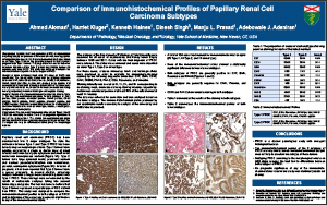
Results: Results of the immunohistochemical stains are listed in Table 1. The best immunohistochemical profile for both subtypes is: CK7: positive, EMA: positive, racemase: positive, PAX-8: positive, CD10: variable, PAX-2: variable, CAIX: negative, vimentin: negative and CK903: negative. There was no correlation between the staining pattern and tumor characteristics such as tumor type, location, nuclear grade, and presence of desmoplasia.
Conclusions: The immunohistochemical profiles of both Types 1 and 2 PRCC are essentially the same. There is no prognostic significance to any of the immunostains. The similar immunohistochemical profile confirms that PRCC is one entity with divergent histologic features. The 2 subtypes can only be differentiated based purely on morphologic features.
©2013 Yale Department of Pathology. All rights reserved.
Any redistribution or reproduction of part or all of the contents in any form is prohibited. You may not, except with express written permission of the author or the Department of Pathology, distribute or commercially exploit the content, nor may you transmit it or store it in any other website or other form of electronic retrieval system, including use for educational purposes.