Can Tall Cell Microcarcinoma be Diagnosed on Fine Needle Aspiration? An Analysis of Clinicopathological Features, Preoperative Fine Needle Aspiration and Genetic Alteration
Jane Bernstein, Manju L. Prasad, David Chhieng, Pei Hui, Adebowale J. Adeniran
Department of Pathology, Yale School of Medicine, New Haven, CT, USA
ABSTRACT
Background: Thyroid papillary microcarcinomas are by definition equal to or less than 1.0 cm in size. These are often considered incidental and clinically insignificant. Tall cell variant of papillary thyroid carcinoma is a rare variant that is associated with a more aggressive behavior and often presents at an advanced stage. Recently we described the tall cell variant of papillary thyroid microcarcinoma (microTCV). Despite their small size, microTCV displayed aggressive pathological features. This study was designed to determine if microTCV have features sufficiently characteristic for their preoperative diagnosis as this would help to plan an appropriate surgical strategy.
Design: microTCV cases over the last 10 years were identified from our files. The cytology slides were reviewed and the findings were correlated with clinicopathologic features and with results of BRAF V600E mutation analysis.
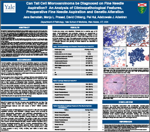
Results: Twenty-one cases were identified (18 females and 3 males). The average age of patients was 53 years (range, 34-73 years). All patients underwent total thyroidectomy. Thirteen of the thyroids contained multifocal microTCV. Tumor size ranged from 0.2 cm to 0.9 cm (mean, 0.70 cm). Background lymphocytic thyroiditis was present in 10 cases. At presentation 29% had lymph node metastasis, vascular invasion was present in 19% while extrathyroidal extension was present in 38% of cases. Fifty eight percent of cases had smears with papillary groups while 68% showed cohesive flat sheets. The frequency of cytologic features were as follows: nuclear grooves – 90%; abundant dense eosinophilic cytoplasm – 90%; nuclear enlargement – 74%; nuclear pseudoinclusions – 74%; irregular nuclear membranes – 47%; powdery chromatin – 42%; crowding and overlapping – 42%. Twenty cases (95%) were positive for BRAF V600E mutation.
Conclusions: microTCV is frequently associated with multifocality, extrathyroidal extension, and lymph node involvement at presentation and high BRAF mutation rate, hence the need for recognition on FNA. Features with high diagnostic yield include cohesive flat sheets, abundant dense eosinophilic cytoplasm, nuclear enlargement and grooves, among others.
©2013 Yale Department of Pathology. All rights reserved.
Any redistribution or reproduction of part or all of the contents in any form is prohibited. You may not, except with express written permission of the author or the Department of Pathology, distribute or commercially exploit the content, nor may you transmit it or store it in any other website or other form of electronic retrieval system, including use for educational purposes.