Development of the Fetal Uterus and Comparison to the Post-Menarchal Uterus
Christopher Flynn, Raffaella A. Morotti, and Fattaneh A. Tavassoli
Department of Pathology, Yale School of Medicine, New Haven, CT, USA
ABSTRACT
Introduction: The in utero phase of uterine development is not fully understood. While it is known that the uterus develops from fusion of the Mullerian ducts, the morphologic evolution post-fusion is not well established. Beyond simple scientific curiosity, understanding the development of various components of the fetal uterus could potentially enhance our understanding of pathologic disorders.
Design: A total of 41 fetal uteri from autopsies performed at our institution between 2004 and 2010 were included in the study. The fetuses ranged in age from 19 to 37 weeks. Each uterus with attached adnexa was sectioned along their long axis to dis-play the full thickness of the uterine wall. The thickness of the endometrium (surface epithelium and stroma) was compared to the myometrial thickness. Various immuno-histochemical stains were performed on the uteri to confirm stromal and myometrial thickness and identify potential proteins/pathways involved in uterine development.
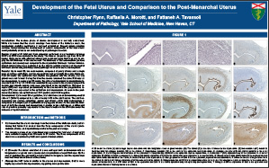
Results: Up to week 20, the endometrium, composed of purely stroma and a single layer of surface epithelium, and the myometrium had essentially the same thickness. Past 20 weeks the epithelium had shallow invaginations into the stroma, but true glands were not formed. During this time the stroma remained the same thickness as the myometrium. In cases over 30 weeks, the ratio of endometrium to myometrium decreased from 1:1 to 1:4. CD10 was consistently expressed in the uterine stroma while desmin and smooth muscle actin were expressed in the myometrium. Estrogen receptor (ER) was expressed predominantly in the stroma while the progesterone receptor (PR) was expressed in the epithelium and myometrium. As seen in the post-menarchal uterus, the epithelium was CK7 positive and CK20 negative.
Conclusions: Up to week 30 of gestation, fetal uteri have an endometrial/myometrial ratio of 1 (E/M=1) compared to a ratio of nearly 1/10 in the adult uterus. The adult endometrium has surface epithelium, glands and stroma, while fetal endometrium is composed of a single layer of epithelium overlying pure stroma. As the absolute thickness of both the stroma and myometrium is greater in the adult uterus, a differential rate of growth is probably responsible for the transformation in the E/M ratio under the influence of cyclic hormones.
©2013 Yale Department of Pathology. All rights reserved.
Any redistribution or reproduction of part or all of the contents in any form is prohibited. You may not, except with express written permission of the author or the Department of Pathology, distribute or commercially exploit the content, nor may you transmit it or store it in any other website or other form of electronic retrieval system, including use for educational purposes.