Significance of Myoepithelial Cell Layer Absence Around Ducts with Papillary Ductal Intraepithelial Neoplasia 1-3/Papillary Ductal Carcinoma In Situ 1-3
Malini Harigopal, Christopher Flynn, Priyanka Karam, Maria Orsaria, Fattaneh A. Tavassoli
Department of Pathology, Yale School of Medicine, New Haven, CT, USA
ABSTRACT
Introduction: While persistence with adjacent conventional invasive carcinoma (IDC), the size and stage of the carcinoma could vary substantially depending on whether the papillary component is interpreted as invasive or intraepithelial.
Design: In a retrospective search of the pathology database at our institution for all PDIN 1-3 (Papillary DCIS) with excisional biopsy between 2001 to 2011, a total of 50 (PDIN, solid), 2 (PDIN, intracystic), 12 PDIN with associated IDC were identified and reviewed. 27 low grade PDIN of solid and intracystic types with or without associated IDC were further evaluated with 3 ME (p63, calponin and CD10) and 2 basement membrane markers (collagen IV & laminin).
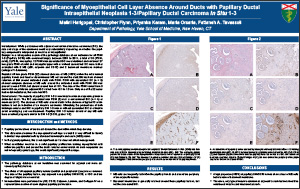
Results: All low grade PDIN (22) showed absence of ME (>90%) within the intra-luminal papillary fronds and focal or discontinuous ME cell around the duct; BM markers showed retention of BM around distended ducts with PDIN. PDIN with associated IDC (n = 5) showed complete absence of ME cells around the distended ducts with PDIN. BM was present around PDIN, but absent around foci of IDC. The size of the PDIN varied from 3 mm to 6.0 cm, while the adjacent IDC varied from <0.1 to 1.3 cm. Only one of the 22 cases had nodal metastases, this case had IDC.
Conclusions: The majority of papillary DIN 1-3 is non-invasive with an expansile growth in distended ducts. The IDC associated with PDIN (5) was a conventional IDC (n = 4) or mucinous (n=1). The absence of ME cells around ducts in the absence of typical IC architecture is not an indication of an invasive carcinoma. Ultimately, the prudent use of both histologic criteria and IHC in papillary DIN 1-3 with or without associated IDC is critical to avoid overstaging and overtreatment. Papillary DIN 1-3 lesions devoid of any ME cells have excellent prognosis similar to DIN 1-3 (DCIS, grades 1-3).
©2013 Yale Department of Pathology. All rights reserved.
Any redistribution or reproduction of part or all of the contents in any form is prohibited. You may not, except with express written permission of the author or the Department of Pathology, distribute or commercially exploit the content, nor may you transmit it or store it in any other website or other form of electronic retrieval system, including use for educational purposes.