HHV-6 Positive Reed-Sternberg Cells in Nodular Sclerosis Hodgkin Lymphoma
Alexa J. Siddon, MD1, Larissa Lozovatsky1, Ayman Mohamed2, and S. David Hudnall, MD1
Department of 1Pathology, Yale School of Medicine, and 2 Precipio Diagnostics, New Haven, CT, USA
ABSTRACT
Classical Hodgkin lymphoma (HL) is comprised of malignant Reed-Sternberg (RS) cells scattered within a mixed inflammatory background. The unusual bimodal age distribution of HL suggests that infectious agents may play a role in etiology. The presence of Epstein-Barr virus (EBV) within the RS cells in a proportion of HL cases supports this idea. However, the most common subtype of HL, nodular sclerosis HL (NSHL), is the subtype least often EBV-associated.
HHV-6 is a near-ubiquitous herpesvirus first acquired in childhood characterized most often by asymptomatic life-long persistence. Since a few reports have suggested a role for HHV-6 in HL, we have analyzed a cohort of NSHL cases for both EBV and HHV-6, and sought to specifically localize these viruses to the malignant RS cells.
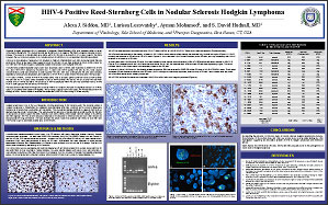
Formalin-fixed paraffin-embedded lymph nodes from 20 primary cases of NSHL were examined by EBER ISH, HHV-6 IHC, and HHV-6 PCR followed by Southern blot. In cases with HHV-6 positive RS cells by IHC, laser capture microdissection (LCM) was performed to collect purified RS cells. DNA from LCM-captured RS cells was extracted, amplified by whole genome amplication, and subjected to HHV-6 PCR to confirm that the RS cells were HHV-6 positive.
Of 17 cases of NSHL, 13 were HHV-6 PCR positive (76%). Five of 20 cases (25%) contained numerous EBV positive RS cells and 10 of 21 cases (48%) contained numerous HHV-6 positive RS cells by IHC (in 3 cases RS cells were positive for both HHV-6 and EBV). The presence of HHV-6 specifically within RS cells was confirmed both by HHV-6 PCR on LCM-captured RS cells and by HHV-6 FISH.
We have demonstrated that HHV-6 genome is present within the neoplastic RS cells of a significant proportion of NSHL cases, most of which were EBER ISH negative. These findings support that in some cases, HHV-6 may play a role in the etiology of NSHL. Further studies to examine the contribution of HHV-6 to the etiology of HL are ongoing.
©2012 Yale Department of Pathology. All rights reserved.
Any redistribution or reproduction of part or all of the contents in any form is prohibited. You may not, except with express written permission of the author or the Department of Pathology, distribute or commercially exploit the content, nor may you transmit it or store it in any other website or other form of electronic retrieval system, including use for educational purposes.