Automated Objective Determination of Percentage of Malignant Nuclei for Mutation Testing
Hollis Viray1, Madeline Coulter1, Kevin Li1, Kristin Lane2, Aruna Madan1, Kurt Schalper1, Kisha Mitchell1, Clifford Hoyt2, and David L. Rimm1
1Department of Pathology, Yale University School of Medicine, New Haven, CT; 2Caliper Life Sciences, Hopkinton, MA, USA
ABSTRACT
Background:
DNA mutations detected in tumors are a critical companion diagnostic test for some new targeted therapies. The accuracy of mutation detection depends on the sensitivity of the assay and on the percentage of tumor cells in the sample. Currently, the malignant cell percentage is judged by eye resulting in a large variation of estimated percentages. This aspect of DNA mutation testing can potentially be standardized by developing a computer algorithm capable of objectively determining the percentage of malignant nuclei in an image of tumor tissue.
Methods:
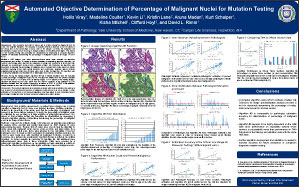
H&E images from colon adenocarcinoma cases were selected for algorithm development and testing. To create a criterion standard for evaluating algorithm accuracy, the nuclei in each image were classified as malignant or benign and counted by a technician, then reviewed by a pathologist. Using inForm software (Caliper Life Sciences), an algorithm was developed to calculate the percentage of malignant cells in a single field of view based on feature extraction involving tissue stain optical densities and morphology. Example regions defining malignant and benign nuclei from 25 cases were used to train the algorithm. The algorithm was subsequently validated on a separate set of 100 images from a tissue microarray.
Results:
Among the training images, Algorithm #9 had a median deviation from the manually counted percentage of malignant nuclei of 5.4%. The algorithm differed from the criterion standard by less than 5.0% on 11 (44.0%) of the 25 training images. For 17 (68.0%) of the training images, Algorithm #9 differed by less than 10.0% from the criterion standard. In the validation set, the algorithm deviated from the criterion standard by a median of 6.2%. 47 (47.0%) of the validation images deviated by less than 5.0% and 58 (58.0%) deviated by less than 10.0%.
Conclusion:
This method represents an exploratory example with future potential to be used as a tool to assist in determining the percent of malignant nuclei present in a tissue sample. Further validation of this algorithm or an improved algorithm may have value to more accurately assess percentage of malignant cells for future companion diagnostic mutation testing.
©2012 Yale Department of Pathology. All rights reserved.
Any redistribution or reproduction of part or all of the contents in any form is prohibited. You may not, except with express written permission of the author or the Department of Pathology, distribute or commercially exploit the content, nor may you transmit it or store it in any other website or other form of electronic retrieval system, including use for educational purposes.