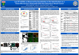
Loss of miR34a as Measured by Quantitative In Situ Hybridization on a Tissue Microarray is Associated with Poor Outcome in Breast Cancer
Seema Agarwal, Jason A. Hanna, and David L. Rimm
Department of Pathology, Yale School of Medicine, New Haven, CT
ABSTRACT (updated)
Introduction:
MicroRNAs (miRNAs) have emerged as key regulators in the pathogenesis of cancers as either oncogenes or tumor suppressors. It has been shown that the loss of miR34a leads to tumor progression and metastasis using cell lines and a limited number of patient samples by RT-PCR. Lack of a robust, reproducible and quantitative method has limited large scale analysis and assessment of miR34a as tumor suppressor marker in cancer. Herein, we have developed and validated a method for the quan-titative analysis of miRNA expression by in situ hybridization (qISH) allowing for the direct assessment of tumor epithelial expression of miR34a in breast cancer.
Method:
Expression level of miR34a was measured in a retrospective breast cancer (n=461) cohort with more than 20 year follow-up using quantitative immunofluorescence (AQUA) technology for ISH in a tissue microarray (TMA) format. The assay was performed in two-fold redundancy with a 40 case index TMA for reproducibility and standardization. Averaged AQUA scores for miR34a were correlated with clinical and pathological characteristics and 20 year disease-free survival in this cohort. An independent breast cancer cohort (n=279) was used as a validation cohort.
Results:
Since miR34a is a tumor suppressor, we determined the threshold at which no specific hybridization was seen (AQUA score =24.0). Repro-ducibility between two different builds (cores) of the TMA was R2 = 0.59. Using overall survival as an endpoint in Kaplan Meier analysis with the threshold of expression as the cutpoint, the group with loss of miR34a had significantly worse survival in training cohort (log rank p = 0.0188) and in validation cohort (log rank p = 0.0024). Cox multivariate analysis including age, nuclear grade, nodal status, ER, PR and Her2 showed miR34a is an independent marker (p=0.0435 and 0.0452) in both training and validation cohorts respectively.
Conclusions:
The microRNA miR34a has been proposed to be a tumor suppressor using mechanistic and functional data. This result, showing loss of miR34a is associated with worse outcome in a large breast cancer cohort provides clinical evidence of the tumor suppressor function of miR34a.
©2012 Yale Department of Pathology. All rights reserved.
Any redistribution or reproduction of part or all of the contents in any form is prohibited. You may not, except with express written permission of the author or the Department of Pathology, distribute or commercially exploit the content, nor may you transmit it or store it in any other website or other form of electronic retrieval system, including use for educational purposes.