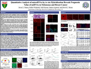
Quantitative Analysis of microRNAs by in situ Hybridization Reveals Prognostic Value of miRNAs in Melanoma and Breast Cancer
Jason A. Hanna, Hallie Wimberly, Salil Kumar, Seema Agarwal, and David L. Rimm
Department of Pathology, Yale School of Medicine, New Haven, CT
ABSTRACT
Introduction:
Most microRNA (miRNA) measurement methods require total RNA extraction which destroys spatial information and makes standardization a challenge. In situ hybridization (ISH) however, allows the direct assessment of miRNA expression levels in tissue and importantly allows for the evaluation of expression in malignant cells as well as stromal cells. We have developed and validated a novel method for the quantitative analysis of miRNA expression by ISH on formalin fixed paraffin embedded tissue microarrays (TMAs) using miR-21, miR-92a, miR-34a, miR-221, miR-205, and Let-7a.
Methods:
In order to quantitatively measure miRNA expression by ISH, DIG labeled LNA modified miRNA probes were employed and multiplexed with DAPI and cytokeratin or S100/GP100 immunofluorescence to apply the AQUA® technology. This quantitative approach allows for the measurement of miRNA expression in subcellular compartments. The assay allows standardization of miRNA measurement and potential for rapid large cohort assessment. We also assessed protein targets of each miRNA by AQUA to investigate the hypothesized inverse relationship between miRNAs and target proteins.
Results:
In hundreds of tissue samples a broad dynamic range and a variable expression pattern specific to each miRNA was observed. Specificity of the assay was validated with anti-miR and miRNA mimic transfected cell lines, miR-21 knock out mouse tissue, and blocking oligo experiments. The assays were performed on near serial sections and were found to have high reproducibility with R2>0.8. miR-221 and miR-205 were found to be prognostic in breast cancer and melanoma respectively (p<0.05). Finally, the inverse relationship between miRNAs and putative targets were not seen in the large population cohorts.
Conclusions:
This specific and reproducible method for the quantitative analysis of miRNA expression provides proof of concept for the use of miRNAs as tissue biomarkers using quantitative ISH. Future studies will further determine the prognostic value of miR-221 and miR-205, and the relationship between miRNAs and target protein expression.
©2012 Yale Department of Pathology. All rights reserved.
Any redistribution or reproduction of part or all of the contents in any form is prohibited. You may not, except with express written permission of the author or the Department of Pathology, distribute or commercially exploit the content, nor may you transmit it or store it in any other website or other form of electronic retrieval system, including use for educational purposes.