Validation of Antibodies to Estrogen Receptor β and Quantitative Assessment of ERβ1 and ERβ5 Expression in Breast Cancer
Hallie Wimberly1, Leigh C. Murphy2, Bruce Haffty3, and David L. Rimm1
1Department of Pathology, Yale School of Medicine, New Haven, CT; 2University of Manitoba, Winnipeg, MB, Canada; 3Robert Wood Johnson Medical School, University of Medicine and Dentistry of New Jersey, Cancer Institute of New Jersey, New Brunswick, NJ
ABSTRACT (updated)
Introduction:
Estrogen receptors are members of the nuclear hormone receptor family that play an important role in breast carcinogenesis and response to endocrine therapy. Though the role of ERα in breast cancer has been studied extensively, little is known about the alternative isoform ERβ. ERβ has significant sequence homology to ERα, but is located on a different chromosome and maintains both overlapping and unique functional attributes. Five variants resulting from alternative splicing of the C-terminal region of ERβ exist. The relevance of ERβ variants in breast cancer outcomes and response to therapy is difficult to assess because of conflicting results in the literature, likely due to variable methods used to assess ERβ in patient tumors.
Methods:
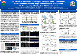
Antibodies against ERβ variants (ERβ1: ThermoScientific PPG5/10; ERβ2/cx: Serotec Clone 57/3; ERβ5: Serotec Clone 5/25) were validated for staining specificity by siRNA knockdown of ESR2 as well as staining reproducibility on FFPE tissue by quantitative immunofluorescence (QIF) using AQUA technology (HistoRx). QIF staining of validated antibodies was then assessed on two separate breast cancer cohorts.
Results:
ERβ1 and ERβ5, but not ERβ2/cx, antibodies were found to be sensitive, specific and reproducible, as shown by reduction in signal after siRNA knockdown in cell lines and reproducible QIF scores on a set of breast cancer control cases. The distribution of both ERβ1 and ERβ5 is similar in two breast cancer cohorts and QIF scores are significantly associated in both cohorts. When patients are stratified into low and high ERβ5 groups based on the median QIF score, high ERβ5 is a trending marker of worse prognosis in (1) ERα positive patients on one of the cohorts examined (p=0.0201) and (2) in patients treated with adjuvant tamoxifen and/or chemotherapy (p=0.0195). ERβ1 does not appear to provide any prognostic information on either cohort.
Conclusions:
Rigorous validation of ERβ antibodies is required for accurate measurement of expression. Assessment of two breast cancer cohorts using validated reagents show that ERβ5, but not ERβ1, is a trending marker of worse prognosis in ERα positive patients and patients treated with adjuvant tamoxifen and/or chemotherapy.
©2012 Yale Department of Pathology. All rights reserved.
Any redistribution or reproduction of part or all of the contents in any form is prohibited. You may not, except with express written permission of the author or the Department of Pathology, distribute or commercially exploit the content, nor may you transmit it or store it in any other website or other form of electronic retrieval system, including use for educational purposes.