This is a core needle liver biopsy (note the shape) that is taken by inserting a needle through the skin into the liver.
The liver demonstrates numerous apoptotic hepatocytes (also called acidophil bodies because of their pink color). This patient was infected with hepatitis C. Virally-infected hepatocytes undergo apoptosis in response to cytotoxic T lymphocytes (CTLs) and thus is an example of extrinsic apoptosis. The process can be activated via the secretion by the CTLs of perforin & granzymes, as well as the binding of Fas on the infected hepatocyte with Fas-ligand on the CTL. In addition to virally-infected cells, CTLs can also attack somatic cells in bone marrow transplant patients (graft-versus-host disease).
The apoptotic cascade involves the activation of caspases (cysteine proteases) which enzymatically degrade structural proteins and activate DNAses, which promotes the characteristic "ladder" cleavage pattern of the cells DNA (lane b).
Intrinsic (a.k.a mitochondrial) apoptosis occurs in many physiologic:
- The involution of hormone-dependent tissues
- Cell loss in proliferating cell populations (germinal centers, lymphomas)
- Elimination of self-reactive lymphocytes
and pathologic conditions:
- Radiation damage to DNA
- Free-radical damage
- Genetic (e.g. accumulation of misfolded protein in the ER)
- Tumors (Bcl-2 is an inhibitor of apoptosis and lymphomas frequently have Bcl-2 mutations that switch it off)
As a routine part of your autopsy, you check the pathology files to determine if there are any pre-mortem pathology specimens from your patient. You find one specimen taken 5 years before the patient death.
Click the button to access the virtual slide

Unable to access?
To observe this specimen click on the button to the left.
What tissue is this?
How was this tissue taken?
What are the scattered eosinophilic (pink) cells? Do they elicit an inflammatory response?
How does this damage compare to what you saw in the kidney? What process is this?
Is this an intrinsic or extrinsic injury, and how is it initiated?
Can you name some of the enzymes involved in this process?
Could this process contribute to the patient's current liver findings?
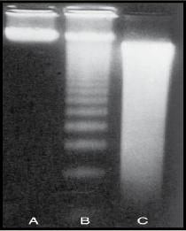
To the left are the DNA profiles of three cells. If you could analyze the DNA from the eosinophilic cells in your case, which profile would they most look like (A, B, or C)?
What other things cause this phenomena?