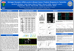
Estrogen Receptor mRNA Levels in Breast Cancer Predicts Response to Tamoxifen
Jennifer M. Bordeaux1, Huan Cheng1, Allison W. Welsh1, Bruce G. Haffty1, Donald R. Lannin1, Xingyong Wu2, Nan Su2, Xiao-Jun Ma2, Yuling Luo2 and David L. Rimm1
1Department of Pathology, Yale University School of Medicine, New Haven, CT; 2Advanced Cell Diagnostics, Hayward, CA, USA
ABSTRACT
Purpose:
Quantification of mRNA has historically been done by reverse transcription polymerase chain reaction (RT-PCR). Recently, a robust method of detection of mRNA utilizing in situ hybridization has been described that is linear and shows high specificity with low background. Here we describe the use of the AQUA method of quantitative immunofluorescence (QIF) for measuring mRNA in situ using ESR1 (the estrogen receptor alpha gene) in breast cancer to determine its predictive value compared to Estrogen Receptor a (ER) protein.
Methods:
Messenger RNA for ER (ESR1) and Ubiquitin C (UbC) were visualized using RNAscope probes and levels were quantified by quantitative in situ hybridization (qISH) on two Yale breast cancer cohorts on tissue microarrays. ESR1 levels were compared to ER protein levels measured by QIF using the SP1 antibody.
Results:
ESR1 mRNA is reproducibly and specifically measurable by qISH on tissue collected from 1993 or later. ESR1 levels were correlated to ER protein levels in a non-linear manner on two Yale cohorts. High levels of ESR1 were found to be predictive of response to tamoxifin.
Conclusion:
Quantification of mRNA using qISH may allow assessment of large cohorts with minimal formalin fixed, paraffin embedded tissue. Exploratory data using this method suggests that measurement of ESR1 mRNA levels may be predictive of response to endocrine therapy in a manner that is different from the predictive value of ER. Further studies are underway using tissue microarrays from other institutions to determine how tissue age affects this assay.
©2012 Yale Department of Pathology. All rights reserved.
Any redistribution or reproduction of part or all of the contents in any form is prohibited. You may not, except with express written permission of the author or the Department of Pathology, distribute or commercially exploit the content, nor may you transmit it or store it in any other website or other form of electronic retrieval system, including use for educational purposes.