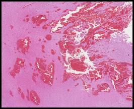
The histology shows liquefactive necrosis surrounded by reactive astrocytes and macrophages filled with myelin breakdown products
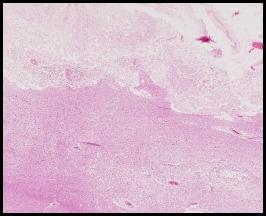
Here are the corresponding microscopic images. What type of necrosis do you see?
Compare the pathology to the normal. Where is the necrosis in the image on the left? How does it differ from that seen in the top image?
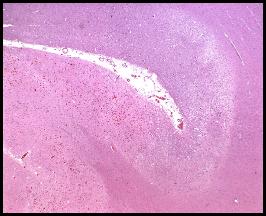
Is there necrosis in this image. If not why not? Would you ever see necrosis in this type of stroke?
In this case, the patient died of cerebral compression due to the expanding hematoma prior to the development of frank necrosis. Had the patient survived, necrosis is clearly possible in areas that infarcted due to the compressive effects.
The necrosis here is less advanced than with the thrombotic infarct. Note the location at the periphery of the brain at the junction of the white and grey matter.